Hello, in this particular article you will provide several interesting pictures of figure 3 from single particle cryo electron. We found many exciting and extraordinary figure 3 from single particle cryo electron pictures that can be tips, input and information intended for you. In addition to be able to the figure 3 from single particle cryo electron main picture, we also collect some other related images. Find typically the latest and best figure 3 from single particle cryo electron images here that many of us get selected from plenty of other images.
 Figure 3 from Single-particle cryo-electron microscopy of We all hope you can get actually looking for concerning figure 3 from single particle cryo electron here. There is usually a large selection involving interesting image ideas that will can provide information in order to you. You can get the pictures here regarding free and save these people to be used because reference material or employed as collection images with regard to personal use. Our imaginative team provides large dimensions images with high image resolution or HD.
 Plasmodium falciparum cryo electron microscopy (cryoEM) protein figure 3 from single particle cryo electron - To discover the image more plainly in this article, you are able to click on the preferred image to look at the photo in its original sizing or in full. A person can also see the figure 3 from single particle cryo electron image gallery that we all get prepared to locate the image you are interested in.
 | Single particle cryo-EM structure of A2AR-BRIL a, b and c, Three We all provide many pictures associated with figure 3 from single particle cryo electron because our site is targeted on articles or articles relevant to figure 3 from single particle cryo electron. Please check out our latest article upon the side if a person don't get the figure 3 from single particle cryo electron picture you are looking regarding. There are various keywords related in order to and relevant to figure 3 from single particle cryo electron below that you can surf our main page or even homepage.
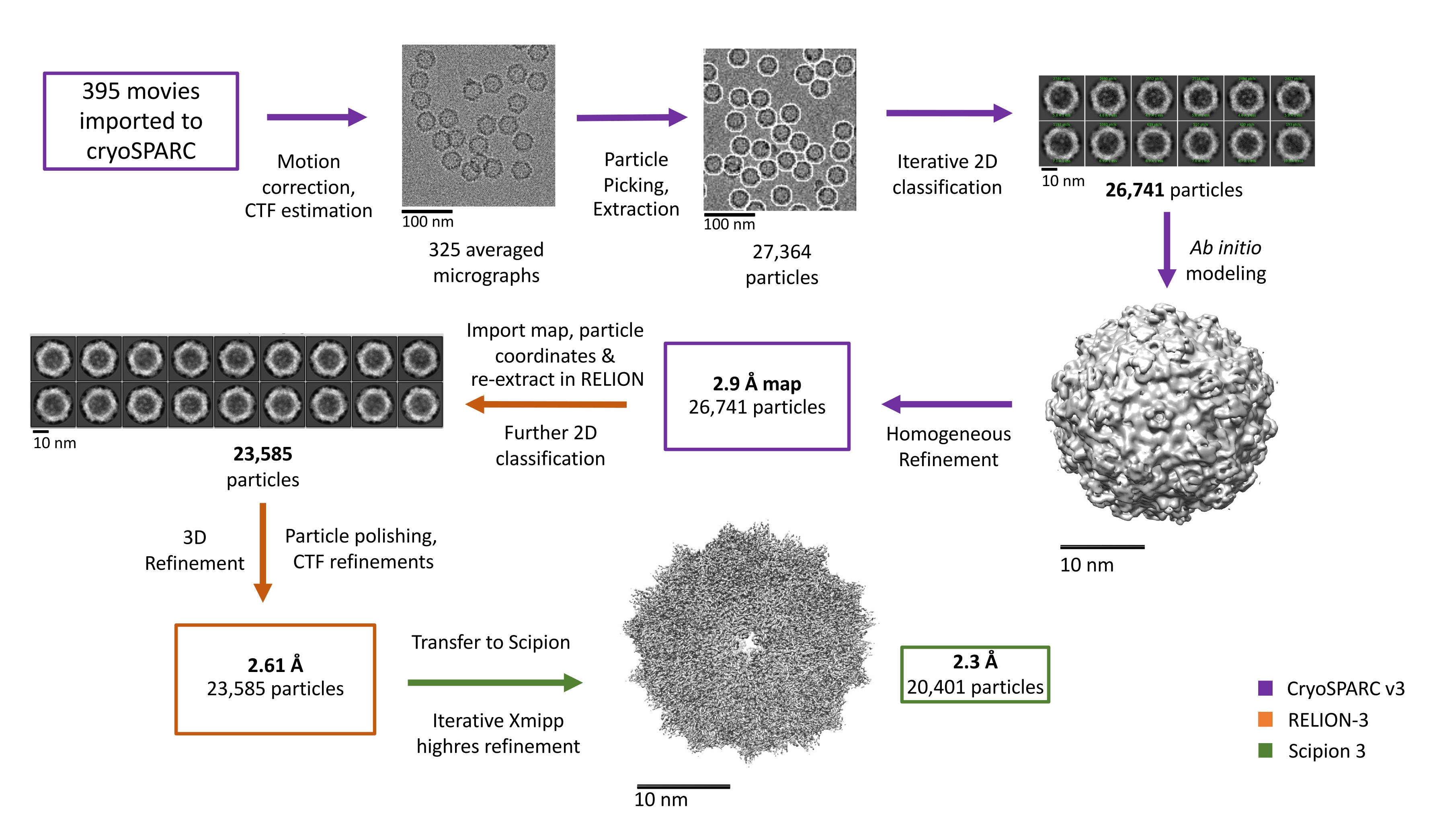 A Robust Single-Particle Cryo-Electron Microscopy (cryo-EM) Processing Hopefully you discover the image you happen to be looking for and all of us hope you want the figure 3 from single particle cryo electron images which can be here, therefore that maybe they may be a great inspiration or ideas throughout the future.
 Simplified single particle cryo-EM data analysis workflow Each step in All figure 3 from single particle cryo electron images that we provide in this article are usually sourced from the net, so if you get images with copyright concerns, please send your record on the contact webpage. Likewise with problematic or perhaps damaged image links or perhaps images that don't seem, then you could report this also. We certainly have provided a type for you to fill in.
 Single-particle cryo-EM structure of a voltage-activated potassium The pictures related to be able to figure 3 from single particle cryo electron in the following paragraphs, hopefully they will can be useful and will increase your knowledge. Appreciate you for making the effort to be able to visit our website and even read our articles. Cya ~.
 Seeing Atoms by Single-Particle Cryo-EM: Trends in Biochemical Sciences Seeing Atoms by Single-Particle Cryo-EM: Trends in Biochemical Sciences
 Single-particle cryo-EM—How did it get here and where will it go | Science Single-particle cryo-EM—How did it get here and where will it go | Science
 Single-particle cryo-EM analysis of human α1β3γ2L GABAA receptor bound Single-particle cryo-EM analysis of human α1β3γ2L GABAA receptor bound
 Cryo-EM single particle analysis with the Volta phase plate | eLife Cryo-EM single particle analysis with the Volta phase plate | eLife
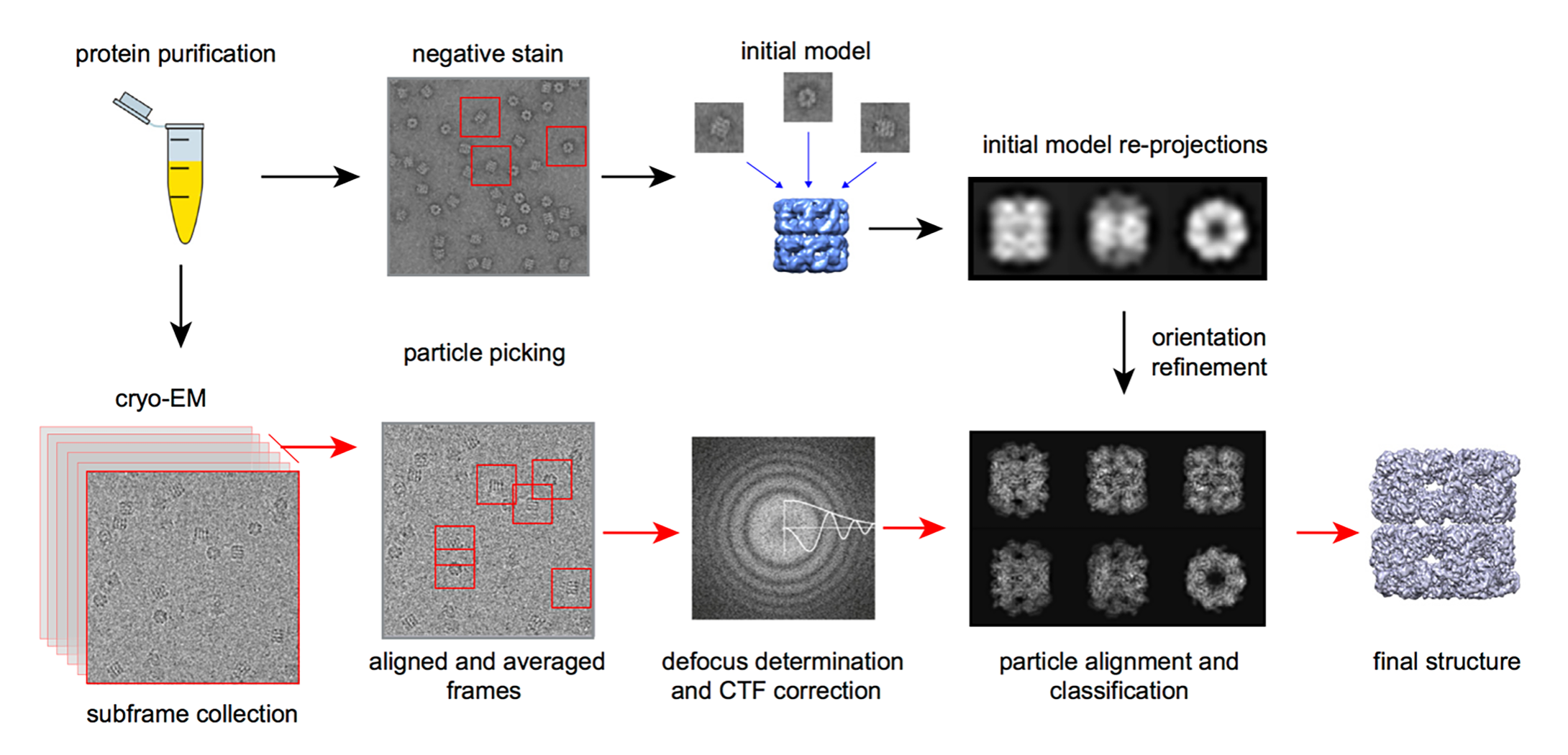 Protein Structure Analysis Using Single Particle Cryo-EM for Protein Structure Analysis Using Single Particle Cryo-EM for
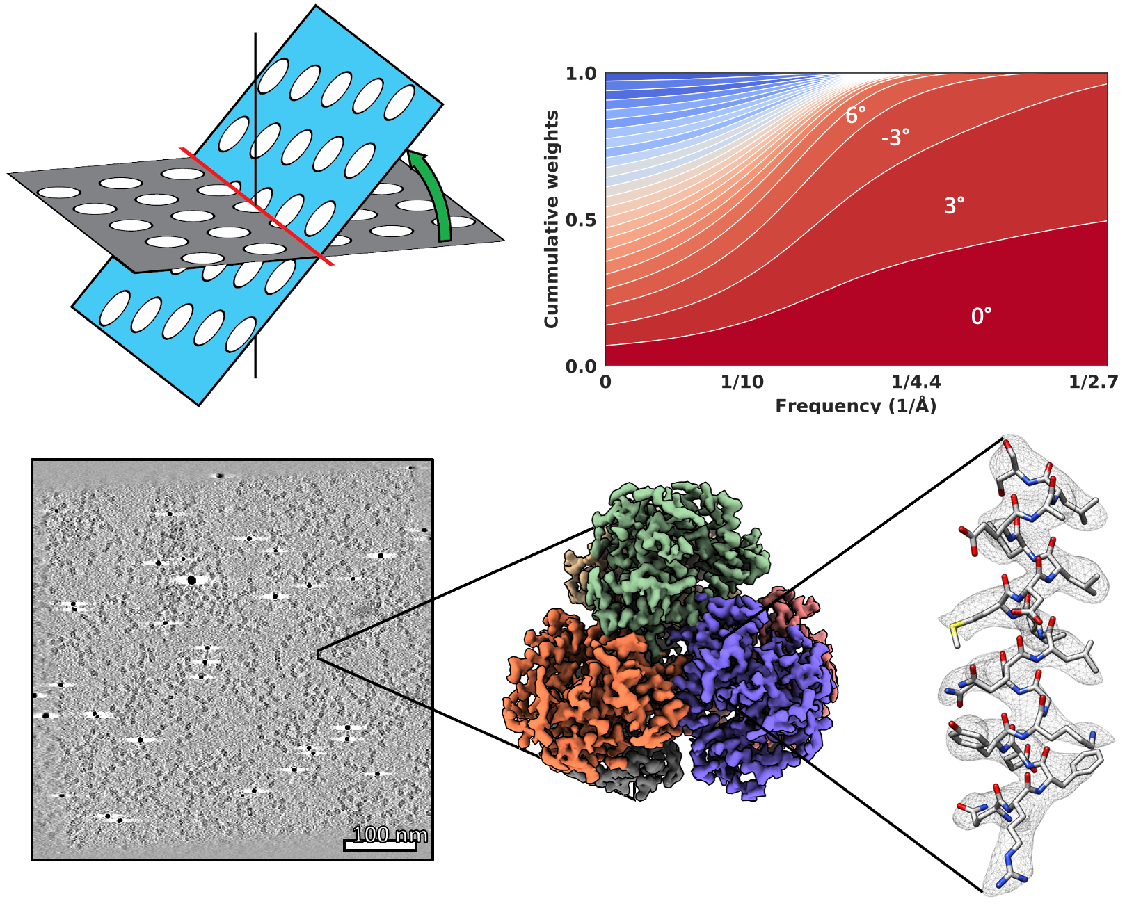 Beam image-shift accelerated data acquisition for near-atomic Beam image-shift accelerated data acquisition for near-atomic
 Single-particle cryo-EM at atomic resolution | bioRxiv Single-particle cryo-EM at atomic resolution | bioRxiv
 Single-particle cryo-EM at atomic resolution | bioRxiv Single-particle cryo-EM at atomic resolution | bioRxiv
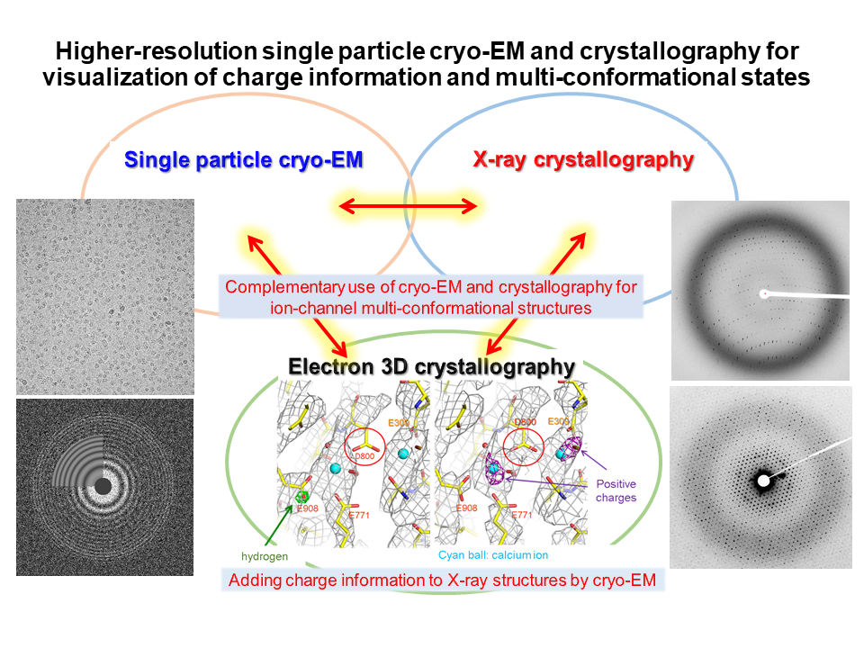 研究内容 - 理化学研究所・独創的研究課題 動的構造生物学 研究内容 - 理化学研究所・独創的研究課題 動的構造生物学
 Single-particle cryo-EM analysis of TDR complex at 35 Å resolution a Single-particle cryo-EM analysis of TDR complex at 35 Å resolution a
 ZPP cryo-EM map of r-EhV-ATPase at 173 Å resolution (a) The cryo-EM ZPP cryo-EM map of r-EhV-ATPase at 173 Å resolution (a) The cryo-EM
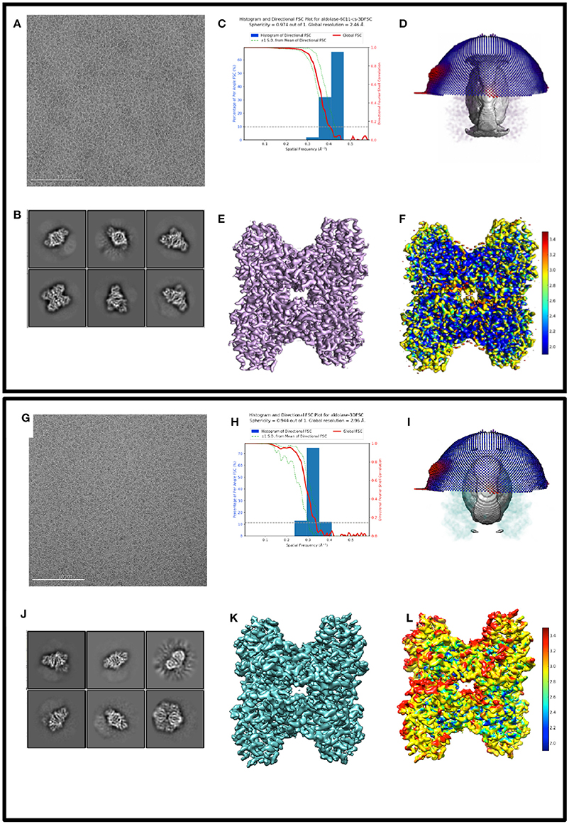 Frontiers | Benchmarking cryo-EM Single Particle Analysis Workflow Frontiers | Benchmarking cryo-EM Single Particle Analysis Workflow
 Routine single particle CryoEM sample and grid characterization by Routine single particle CryoEM sample and grid characterization by
 Schematics of the single-particle cryo-EM processing workflow (A Schematics of the single-particle cryo-EM processing workflow (A
 Single-particle cryo-EM at atomic resolution | bioRxiv Single-particle cryo-EM at atomic resolution | bioRxiv
 Single-particle cryo-EM at atomic resolution | bioRxiv Single-particle cryo-EM at atomic resolution | bioRxiv
 Cryo-electron Microscopy Single-Particle Analysis - Nanomega BioAI Cryo-electron Microscopy Single-Particle Analysis - Nanomega BioAI
 Single-particle cryo-EM structure of the ²⁴⁷DLIIKGISVHI²⁵⁷ fibril Single-particle cryo-EM structure of the ²⁴⁷DLIIKGISVHI²⁵⁷ fibril
 Single-particle cryo-EM analysis of the uniplex in high-Ca² Single-particle cryo-EM analysis of the uniplex in high-Ca²
 Figure 2 from Single-particle cryo-electron microscopy of Figure 2 from Single-particle cryo-electron microscopy of
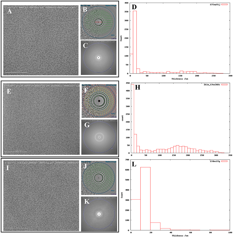 Frontiers | Benchmarking cryo-EM Single Particle Analysis Workflow Frontiers | Benchmarking cryo-EM Single Particle Analysis Workflow
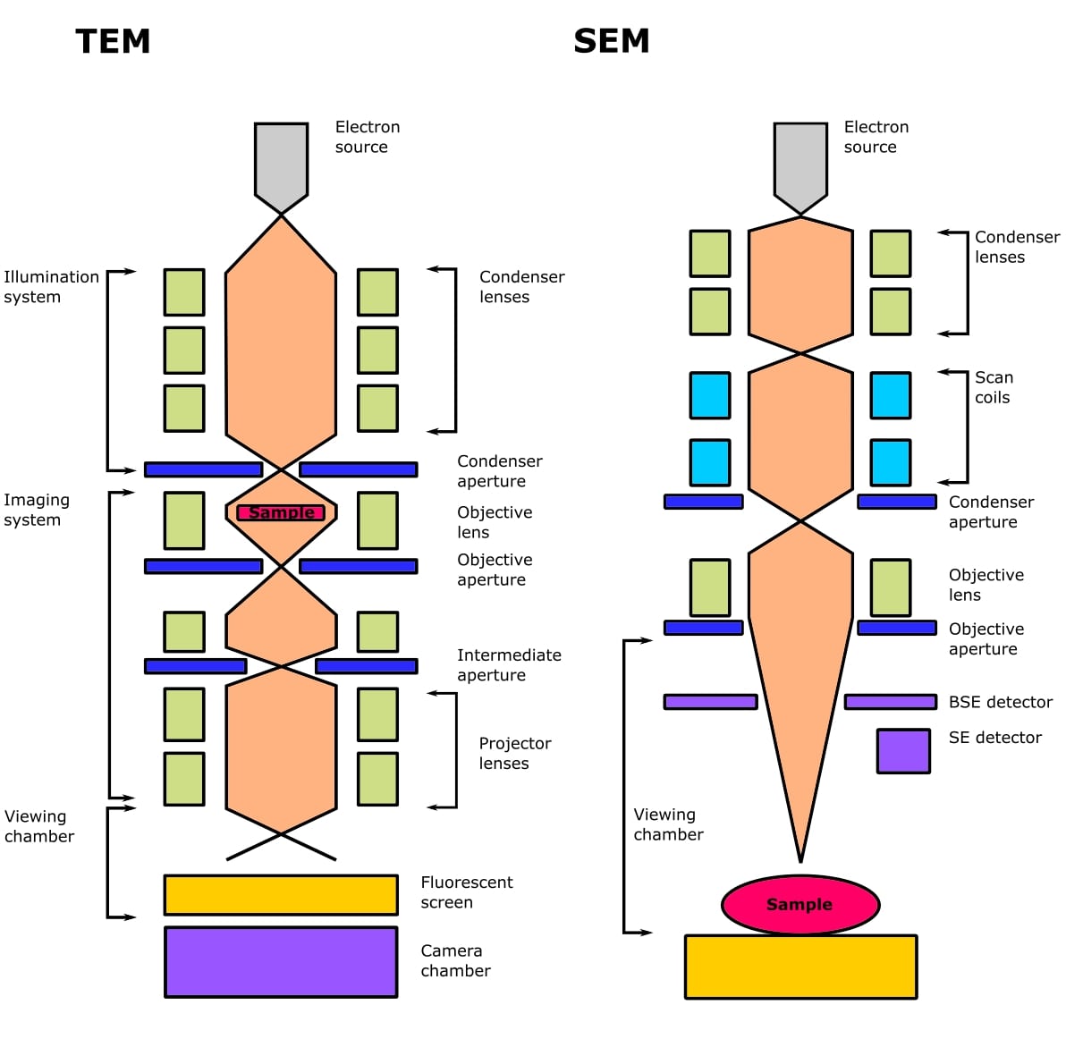 What is Cryo-Electron Microscopy? Discover the fundamentals What is Cryo-Electron Microscopy? Discover the fundamentals
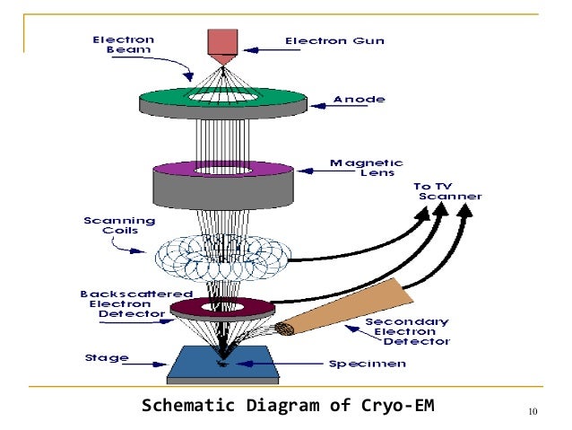 Cryo electron microscopy Cryo electron microscopy
 The cryo-electron microscopic structure of the ELYSC-nucleosome The cryo-electron microscopic structure of the ELYSC-nucleosome

